Disturbo muscoloscheletrico BMC.2014 ago 11;15:271. doi: 10.1186/1471-2474-15-271.
Qian Wang ,
Wenchao Wu ,
Xiaoyu Han ,
Ai Zheng ,
Canzone Lei ,
Jiang Wu ,
Huaiqing Chen ,
Chengqi Lui ,
Fengming Luo ,
Xiaojing Liu 1 Affiliazione
- 1Centro di Medicina Riabilitativa, Ospedale della Cina Occidentale, Università di Sichuan, Chengdu, Sichuan, P, R, Cina. xiaojingliu67@gmail.com.
Astratto
Sfondo: il campo elettromagnetico pulsato (PEMF) è una terapia fisica non invasiva utilizzata nel trattamento della pseudoartrosi di frattura o della guarigione ritardata. Il PEMF può facilitare la differenziazione osteogenica delle cellule staminali mesenchimali del midollo osseo in vitro. Le cellule epiteliali amniotiche (AEC) sono state proposte come potenziale fonte di cellule staminali per la terapia cellulare. Tuttavia, non è noto se il PEMF possa modulare la differenziazione osteogenica degli AEC. Nel presente studio sono stati studiati gli effetti della PEMF sulla differenziazione osteogenica degli AEC.
Metodi: Gli AEC sono stati isolati dalla membrana amniotica della placenta umana mediante digestione con tripsina e sono stati indotti da PEMF e/o mezzo di osteoinduzione. Dopo 21 giorni abbiamo utilizzato la RT-PCR in tempo reale e l'immunocitochimica per studiare l'espressione dei marcatori degli osteoblasti. La trasduzione del segnale dell'osteogenesi è stata ulteriormente studiata.
Risultati: La stimolazione PEMF, o il solo mezzo di osteoinduzione, potrebbe indurre la differenziazione osteogenica degli AEC, come dimostrato dall'espressione di geni e proteine specifici degli osteoblasti, tra cui la fosfatasi alcalina e l'osteocalcina. Inoltre, una combinazione di PEMF e mezzo di osteoinduzione ha avuto effetti sinergici sulla differenziazione osteogenica. Nel nostro studio, l'espressione genica di BMP-2, Runx2, β-catenina, Nrf2, Keap1 e integrinaβ1 è stata sovraregolata nella differenziazione osteogenica degli AEC indotti da PEMF e/o dal mezzo di osteoinduzione.
Conclusioni: l'applicazione combinata di PEMF e mezzo di osteoinduzione è sinergica per la differenziazione osteogenica degli AEC. Potrebbe essere un nuovo approccio nella medicina rigenerativa ossea.
Figure 1 Characterization and phenotype of isolated Amniotic epithelial cells (AECs). (A) Primary culture of human AECs. AECs were successfully isolated from human term placenta and formed a confluent monolayer of cobblestone-shaped epithelial cells after 3 days of culture in the standard culture media (magnification of 200×).
(B) Surface antigen SSEA-4 and the pluripotency marker Oct-4 in primary cultured AECs. Flow cytometry revealed the presence of SSEA-4 (54.2%) and Oct-4 (92.8%) in primary cultured AECs. The open peaks show the isotype-matched antibody control.
(C) Specific marker of epithelial cells identified by immunocytochemistry. Primary cultured AECs could express cytokeratin 19, the epithelial cell marker (magnification of 200×).
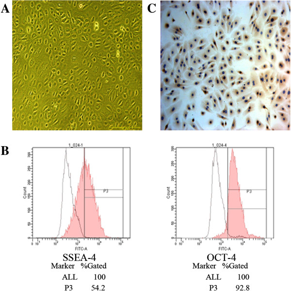 Figure 2 Combined treatment of PEMF and osteo-induction medium increased ALP (A) and OC (B) mRNA expression.
 Experiments were performed three times with the similar results (n = 3 in each group).
* indicates
P < 0.05
vs Control,
# indicates
P < 0.05
vs PEMF and & indicates
P < 0.05
vs OM. Abbreviations: Control, standard culture media; PEMF, PEMF exposure; OM, osteo-induction medium; PEMF + OM, combined treatments with PEMF and osteo-induction medium; ALP, alkaline phosphatase; OC, osteocalcin.
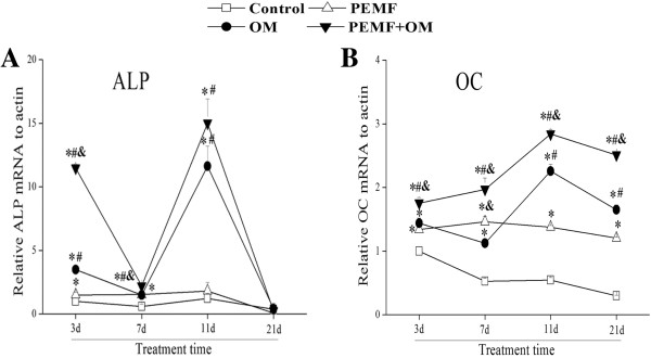 Figure 3 ALP activity was increased during the osteogenic differentiation of AECs. (A)
 The ALP activity was detected by BCIP/NBT staining (magnification of 400×).
(B) The percent of ALP-positive cells (containing blue, insoluble, granular dye deposit) was calculated by ImageJ software Experiments were performed three times with the similar results (n = 3 in each group).
* indicates
P < 0.05
vs Control,
# indicates
P < 0.05
vs PEMF and & indicates
P < 0.05
vs OM.
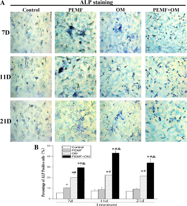 Figure 4 OC protein expression was induced to rise during the osteogenic differentiation of AECs. (A)
 The expression of OC protein was analyzed by immunocytochemistry (magnification of 400×).
(B) The integrated optical density (IOD) of OC was measured by ImageJ software. Experiments were performed three times with the similar results (n = 3 in each group).
* indicates
P < 0.05
vs Control,
# indicates
P < 0.05
vs PEMF and & indicates
P < 0.05
vs OM.
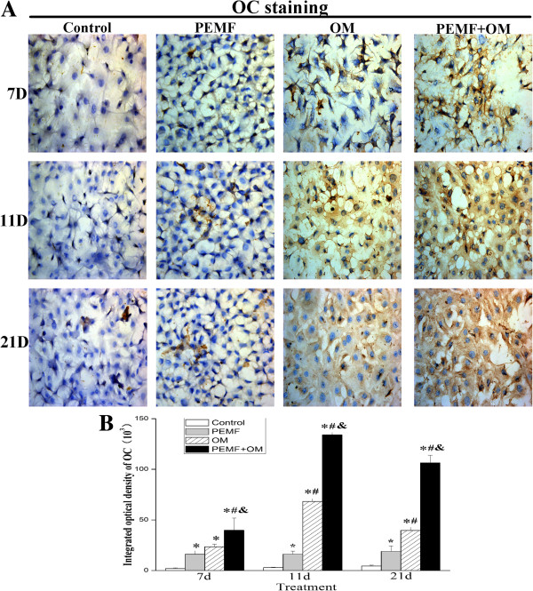 Figure 5 A marked increase of calcium deposits was evoked during the osteogenic differentiation of AECs. (A)
 Calcium deposition was assessed by alizarin red S staining (arrows) (magnification of 400×).
(B) The amount of calcified deposition was semi-quantitatively calculated with ImageJ software. Experiments were performed three times with the similar results (n = 3 in each group).
* indicates
P < 0.05
vs Control,
# indicates
P < 0.05
vs PEMF and & indicates
P < 0.05
vs OM.
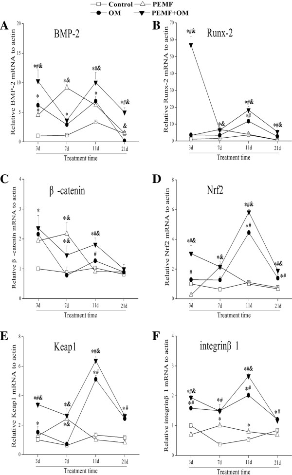
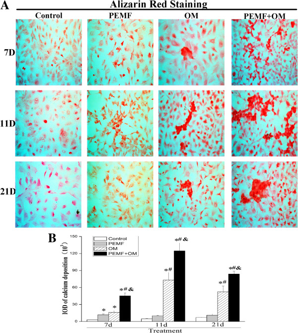 Figure 6 Molecular mechanisms linked to PEMF-induced osteogenic differentiation of AECs.
 Targeted genes involve:
(A) bone morphogenetic protein 2 (BMP-2);
(B) Runx-2;
(C) β-catenin;
(D) Nrf2;
(E) Keap1;
(F) Integrinβ1. Experiments were performed three times with the similar results (n = 3 in each group).
* indicates
P < 0.05
vs Control,
# indicates
P < 0.05
vs PEMF and & indicates
P < 0.05
vs OM.